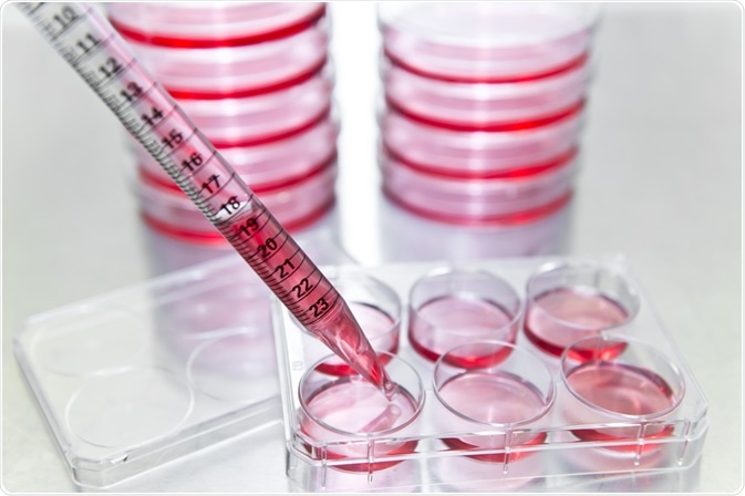
Cell growth and division result in an increase in the number of cells. This is known as cell proliferation. To measure this, cell proliferation assays are utilized. This article will discuss what cell proliferation assays are and why measuring cell proliferation is important.

Image Credit: unoL/Shutterstock.com
What is a cell proliferation assay?
Cell proliferation assays are in vitro techniques. Methods differ in how sensitive they are, their compatibility with high-throughput analysis, and their reproducibility. There are many types of assays used to measure cell proliferation.
Applications for cell proliferation assays include measuring cellular division, DNA synthesis, the number of cells in a sample over time, and metabolic activity within the cell. Researchers choose the assay method depending on the expected outcome of the experiment as well as the type and number of cells within the sample
There are both direct and indirect cell proliferation measurement methods, and these can be performed as both continuous (over time) and endpoint assays. A common conventional method employed is using a hemocytometer to count cell numbers. This is low-cost and direct but relies on large cell counts and specialized training to avoid errors and deviations in the data and is not compatible with high-throughput analysis.
For this reason, multiwell-plate assay techniques were developed. Multiwell-plate assays use luminescence to detect cell numbers and provide data about cell proliferation. The luminescence signal is directly proportional to metabolic activity within the cell.
In recent years, high content imaging platforms have provided new tools to provide crucial phenotypic information on cell populations. These methods, of which there is a growing number in a variety of systems, monitor the proliferation of cells within a sample whilst providing improvements in quantitative and qualitative data collection.
The cell proliferation assay chosen for a study depends on its specific advantages and disadvantages for the reasons listed above (sensitivity, how well it can be reproduced, and compatibility with high-throughput analysis) and the use of a particular assay is considered according to the question researchers aim to answer.
Type of cell proliferation assay
Today, many different types of cell proliferation assays have been developed. They are divided into four main methods: ATP concentration, DNA synthesis, a cell proliferation marker, and metabolic activity assays. Detection methods used include spectrophotometry and Enzyme-linked immunosorbent assays (ELISA.)
- ATP concentration assays – these assays are used to measure the concentration of adenosine triphosphate (ATP) within a cell. As dead cells, or those close to death, contain little ATP, accurate information of cell proliferation can be gathered. These methods are suited to high-throughput analysis. Luminescent biomarkers such as luciferase are used.
- DNA synthesis assays – these assays are used to measure DNA synthesis within a cell population. DNA synthesis assays use radioactively labeled 3H-thymine to detect DNA within cells. Newly proliferated cells incorporate the label, providing extremely sensitive and accurate detection of cell lines. Alternatively, non-radioactive labels such as bromodeoxyuridine can be used.
- Cell proliferation marker assays – This method uses specific monoclonal antibodies to detect antigens that are only present on the surface of proliferating cells. This type of assay is commonly used in cancer research to detect cancerous cells both in vivo and in vitro. However, because this method requires tissue sectioning, it is not compatible with high-throughput analysis. Other markers used include phosphorylated histone H3.
- Metabolic activity assays – Metabolic activity is an accurate marker of cell proliferation. These assays use bioluminescent dyes such as Alamar Blue and Tetrazolium salts to detect lactate dehydrogenase, which increases in activity during proliferation. Each dye has its advantages, and this means that it can be used with a variety of instruments and high-throughput analysis methods.
Each assay has specific advantages and disadvantages. For example, BrdU assays, a common DNA synthesis cell proliferation analysis method, has the advantage of single-cell resolution, but can potentially cause DNA damage.
Live/Dead assays have the advantage of live cell analysis but suffer from background autofluorescence. Trypan Blue can be is low cost but has a degree of inaccuracy. It is therefore important to choose the assay based on these factors.
The importance of measuring cell proliferation
Elucidating information about cell behavior and biochemistry is important for several reasons, including disease research. Diseased cells exhibit different behaviors than normal, healthy cells, including their rates of proliferation. By measuring cell proliferation in different cell populations, information on their disease state and therefore their relative health can be determined.
Another key area of research where cell proliferation is measured is IVF. By measuring proliferation, the effectiveness of IVF treatment and the effects upon developmental behavior can be monitored. Research indicates that embryos experience developmental difficulties if they have lower or higher cell numbers than normal. Cell number is, therefore, a critical factor for the healthy development of the fetus. Abnormal cell proliferation can lead to detrimental health problems for the resultant child.
Measuring this is, therefore, crucial for medical and life sciences researchers. Studies using cell proliferation assay techniques provide data that is used for successful treatments in many branches of clinical science. They are used for testing growth factors, drug reagents, and analyzing cell activity and cytotoxicity.
Cell proliferation assays – A robust and accurate technology
Cell proliferation assays provide accurate and reliable information on cell numbers, growth, and proliferation. Each assay has its own advantages and disadvantages, and these factors as well as the question researchers are trying to answer in the study must be carefully considered before using a particular assay.
References:
- Kong, X et al. (2016) The Relationship between Cell Number, Division Behavior and developmental Potential of Cleavage Stage Human Embryos: A Time-Lapse Study PLoS One 11(4): e0153697 [Accessed online 14th September, 2021] https://www.ncbi.nlm.nih.gov/pmc/articles/PMC4831697/
- Sigmaaldrich.com (website) Cell Viability and Proliferation Assays [Accessed online 14th September, 2021] https://www.sigmaaldrich.com/GB/en/technical-documents/technical-article/cell-culture-and-cell-culture-analysis/imaging-analysis-and-live-cell-imaging/cell-viability-and-proliferation
- Adan, A. Kiraz, Y & Baran, Y (2016) Cell Proliferation and Cytotoxicity Assays Curr Pharm Biotechnol. 17(4): pp 1213-1221 [Accessed online 14th September, 2021] https://pubmed.ncbi.nlm.nih.gov/27604355/
Further Reading
- All Laboratory Techniques Content
- Electrochemical Analysis
- Life Science Applications of Chromatography
- Thermal Analysis of Pharmaceutical Materials
- Using Dynamic Light Scattering (DLS) for Liposome Size Analysis
Last Updated: Nov 12, 2021

Written by
Reginald Davey
Reg Davey is a freelance copywriter and editor based in Nottingham in the United Kingdom. Writing for News Medical represents the coming together of various interests and fields he has been interested and involved in over the years, including Microbiology, Biomedical Sciences, and Environmental Science.
Source: Read Full Article
