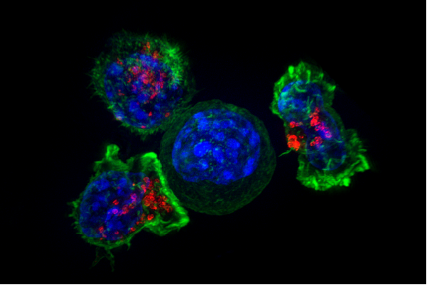
The ability to sequence the genome of a tumor has revolutionized cancer treatment over the last 15 years by identifying drivers of cancer at the molecular level. But understanding the genetic diversity of individual cells within a tumor and how that might impact the disease progression has remained a challenge, due to the current limitations of genomic sequencing.
Using a microfluidic droplet based single cell sequencing method, USC researchers have simultaneously sequenced the genomes of close to 1,500 single cells, revealing genetic diversity previously hidden in a well-studied melanoma cell line.
The study, just published in Nature Communications Biology, demonstrates the ability of single-cell sequencing to reveal possible evolutionary trajectories of cancer cells.
“We used this approach to examine a standard cancer cell-line, examined thousands of times by many different labs,” said David Craig, Ph.D., co- director of the Institute of Translational Genomics at Keck School of Medicine of USC and study author. “What was really surprising here, was with this technology we uncovered complexity we did not expect. This line actually consistently became a mixture of different types of cells. Reexamining decades of prior work on this line—now with this new information—we have new insights into tumor evolution.”
Getting a high-resolution view of cancer’s complexity
Currently, the genetic information of a tumor is typically obtained by sequencing millions of tumor cells together, rather than individually. While this method offers a broad view of the genetic makeup of the tissue it can miss small populations of cancer cells within a tumor that are different from the majority of cells.
With other approaches that analyze the DNA of individual cells, the process is laborious, taking weeks to process just a few cells and requiring resources that most laboratories do not have.
For this study, researchers used an emerging technique called “single-cell copy number profiling.” developed by 10X Genomics with novel analysis methods that integrated these results with those of historical methods.
“Instead of analyzing tissue DNA that is the average of thousands of cells, we analyzed the individual DNA of close to 1500 cells within a single experiment,” said Dr. Enrique Velazquez-Villarreal, lead author and assistant professor of translational genomics at Keck School of Medicine at USC. “Studying cancer at this higher resolution, we can discover information that lower-resolution bulk sequencing misses.”
Their analysis revealed at least four major sub-populations of cells, also known as clones, that are expected to have, at some point during the cancer cell line’s evolution, mutated from the original cancer cell.
The ability to identify sub-clones in cancer tissue could provide important biological insights into how cancer progresses, how it spreads and why it can become resistant to treatment.
“What if there’s a small population of cells in a tumor that has acquired a change that makes them resistant to therapy? If you were to take that tumor and just grind it up and sequence it, you may not see that change,” said John Carpten, Ph.D., study author and Co-Leader of the Translational and Clinical Sciences Program at the USC Norris Comprehensive Cancer Center, Chair of the Department of Translational Genomics, Keck School of Medicine, and Co-Director of the USC Institute for Translational Genomics. “If you go to the single cell level, you not only see it, but you can see the specific population of cells that has actually acquired that change. That could provide earlier access to the molecular information that could help define treatment approaches.”
Source: Read Full Article
