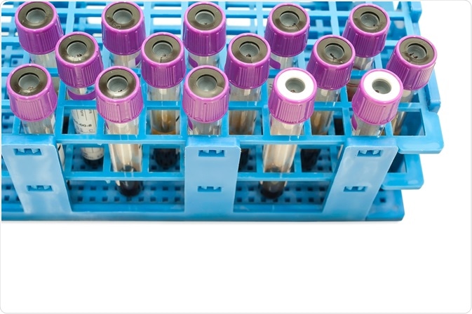
Characterizing tumors can be difficult when they are located inside an organ, often requiring elaborate biopsy procedures to collect tumor tissue. However, tumors release characteristic compounds into the bloodstream that could serve as biomarkers, such as extracellular vesicles (EV) that bud from cells.
 Image Credit: Chansom Pantip / Shutterstock.com
Scientists have recently shown that Imaging Flow Cytometry (IFC) can be used to detect such EVs in blood samples based on specific membrane protein compositions that can be measured with antibody labeling.
The research has been published in the Journal of Extracellular Vesicles.
The identification of tumors in cancer patients is usually carried out by expensive medical imaging examinations along with surgical biopsies. Common imaging procedures include magnetic resonance imaging (MRI) and computed tomographic (CT) reconstruction, where tumors show aberrant structural features.
However, these examinations are expensive and do not allow the specification of the cancer cell type, and thus are insufficient to identify a suitable therapy against the cancer. Following the localization of a tumor by an imaging examination, surgical biopsies are normally carried out to extract a sample from the cancer tissue. From this sample, the specific cancer cell type can be determined by laboratory assays.
With the aim to facilitate cancer screening and to monitor the success of a cancer therapy, attempts have been made previously to detect cancer biomarkers floating in the bloodstream. For instance, cancer cells leave tumor tissues at some frequencies and become circulating tumor cells (CTCs). Tumor-specific, mutated DNA (circulating tumor DNA, ctDNA) has been found in blood samples.
However, it can be difficult to extract CTCs from blood to analyze them in the absence of other blood cells. Floating ctDNA meanwhile is not protected against degradation, and its signal can be overshadowed by cell-free (cf-)DNA from non-cancerous cells.
Researchers with affiliations in Germany, US, Sweden and UK, led by Dr. Franz Lennard Ricklefs and Professor Katrin Lamszus (Hamburg, Germany), have now shown in cell cultures and mouse models that extracellular vesicles (EV) budded from cancer cells can be identified in blood samples by imaging flow cytometry (IFC).
Immunolabeling of extracellular vesicles from tumors
The scientists first analyzed cell cultures of cancer and non-cancer cell lines, where EVs that had been released into the culture liquid can be collected by ultracentrifugation. These samples allowed them to determine the efficiency of tagging the EVs with antibodies linked to fluorescent marker molecules.
They found a good binding of antibodies against the membrane proteins CD9, CD63, and CD81, proteins of the “tetraspanin” family, which are ubiquitous EV markers. With these immunofluorescent labels, the investigators were able to carry out imaging flow cytometry that can detect EVs in liquid samples based on light scattering and specific fluorescence.
After their method development, the scientists demonstrated the application of their method in mouse models after injection of glioma cells, a type of cancer cell line which derives from brain (glial) cells. Additionally, they tested their method with blood samples taken from brain tumor (glioblastoma) patients.
Aberrant membrane protein ratios associated with cancer
The researchers demonstrated that EVs derived from different cancer cell lines were enriched in CD63 and CD81 when compared to the composition of EVs from non-cancer cell lines. The application of imaging flow cytometry (IFC) allowed them to collect multiparametric information from individual EVs based on light scattering and fluorescence.
This appeared of particular importance when the investigators carried out in vivo experiments, where EVs from non-cancerous tissues outnumbered EVs from the cancer cells.
The detection of tumor-specific EVs by IFC represents a promising step towards a liquid biopsy tool to monitor cancer progression in patients. This could become particularly useful during cancer therapies, where frequent monitoring of cancer progression could help clinicians to adjust the therapy at an early stage or identify when a CT is recommended.
Moreover, the identification of tumor-specific EVs in blood samples opens the possibility also to isolate antibody-tagged EVs, thus enabling further molecular characterization of EVs collected from actual blood samples of patients, rather than being derived from in vitro cell cultures.
Further Reading
- All Cancer Content
- What is Cancer?
- What Causes Cancer?
- Cancer Glossary
- Cancer Classification
Last Updated: Nov 11, 2019

Written by
Sally Robertson
Sally has a Bachelor's Degree in Biomedical Sciences (B.Sc.). She is a specialist in reviewing and summarising the latest findings across all areas of medicine covered in major, high-impact, world-leading international medical journals, international press conferences and bulletins from governmental agencies and regulatory bodies. At News-Medical, Sally generates daily news features, life science articles and interview coverage.
Source: Read Full Article
