The Different Types of Immunostaining
Immunostaining encompasses numerous techniques that are suited to a variety of different applications.
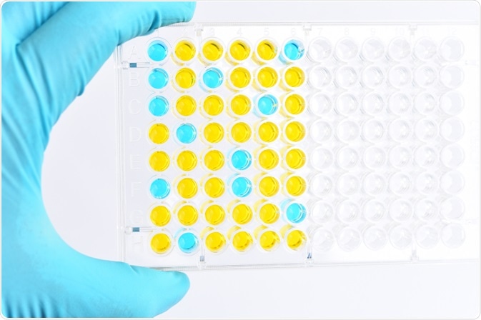
Image Credit: Jarun Ontakrai/Shutterstock.com
However, they are all methods that rely on the use of antibodies to detect and identify proteins within biological samples. They can be used to asses and identify the topographical distribution of abnormal cells, blasts infiltrates, and megakaryocytes.
The term was coined back in 1941 when it was first used to describe immunohistochemical staining. These days, immunohistochemical staining is just one of several established immunostaining techniques, including enzyme-linked immunosorbent assay, flow cytometry, immuno-electron microscopy, and Western blotting.
The techniques are commonplace in biology and molecular biology labs, and are used for a variety of applications in a wide range of fields of study, from oncology to hydrobiology.
Here, we describe the five types of immunostaining techniques.
Enzyme-linked immunosorbent assay (ELISA)
The enzyme-linked immunosorbent assay, also known as ELISA, is commonly used in biochemistry. Developed in 1971 by Engvall and Perlmann, the method quantifies peptides, proteins, antibodies, and hormones present in a sample by immobilizing an antigen on a solid surface before it is complexed with an antibody that is associated with an enzyme.
Identification is then possible when the conjugated enzyme activity is assessed through incubation with a substrate, resulting in a measurable product.
Flow cytometry
When the physical and chemical characteristics of cells or particles are sought to be determined, flow cytometry is often the most suitable method. The technique was established in the 1950s, and over the decade's many advancements have been achieved in its methodology and equipment. Currently, measurements are made from cells in solution while they travel through the instrument’s laser at speeds of 10,000 cells per second. Flow cytometry offers benefits to the technicians who choose to use it, including high sample throughput speed, making it an attractive option as an immunostaining assay.
Immuno-electron microscopy (EM immunolabelling)
Immuno-electron microscopy, also referred to as EM immunolabelling and immuno-EM, is a technique that tags antibody molecules with electron-dense substances, usually, and most effectively, being small gold particles, which are seen during the analysis as easy to spot dark dots. The assay allows for the simultaneous detection of more than one type of molecule because particles of different sizes can be used to tag different antibodies.
The technique was first developed as a diagnostic aid that assisted in the detection and identification of viruses, such as gastroenteritis and rotavirus. Today it is still used to diagnosis a variety of viral infections. It is considered to be one of the most sensitive and rapid methods for this application.
Immunohistochemistry
The most common application of immunostaining is immunohistochemistry, which is used to assist in the diagnosis of various diseases, including different types of cancer. It has also shown its use in neuropathology, and hematopathology, helping in the classification of diseases in these groups and evolving criteria for their diagnosis. Another area in which it has made a significant impact is that of genetic study, where it has been used to determine the role of specific gene products, elucidating their function in vital biological processes. The technique has become invaluable to both medical research and clinical diagnostics.
The method involves selectively identifying antigens in a sample of cells within a tissue section through the principle that certain antibodies will bind to specific antigens present in the tissue. It was established back in the 1930s before it was first reported in 1941.
The initial principle outlined that antibodies labeled with a fluorescent dye could detect pneumococcal antigens in infected tissues. Since then, the technique has been developed, and new enzyme labels have been introduced, including peroxidase, alkaline phosphatase, and colloidal gold. The use of radioactive elements has also been developed for use with autoradiography.
Western blotting
The final immunostaining method is the Western blot method, a widely used technique that has firmly ingrained itself in the fields of cell and molecular biology. Western blot allows researchers to determine and quantify the proteins existent within a cell, identifying specific proteins out of the mixture of proteins that are present in cell samples.
There are three parts to the Western blot method, the first is separation by size, the second being transfer to a solid support, and finally, a target protein is marked using a suitable primary and secondary antibody to visualize it.
Summary
Immunostaining methods have become essential to numerous branches of scientific study, they have also become well established in various clinical applications, mostly in assisting in diagnosis as well as determining characteristics that facilitate more accurate diagnostic criteria.
Since the immunohistochemistry technique was first reported on in 1941, four further types of immunostaining techniques have emerged: enzyme-linked immunosorbent assay, flow cytometry, immuno-electron microscopy, and Western blotting. These methods are being expanded and developed on all the time, growing their use in different applications, and improving on their accuracy and reliability.
Sources:
- Kaliyappan, K., Palanisamy, M., Duraiyan, J. and Govindarajan, R. (2012). Applications of immunohistochemistry. Journal of Pharmacy and Bioallied Sciences, 4(6), p.307. https://www.ncbi.nlm.nih.gov/pmc/articles/PMC3467869/
- Maity, B., Sheff, D. and Fisher, R. (2013). Immunostaining. Laboratory Methods in Cell Biology – Imaging, pp.81-105. https://www.ncbi.nlm.nih.gov/pubmed/23317899
- Saad, A. (2015). Brain Metastasis in Patients with Non-Small Cell Lung Cancer: Immunohistochemical Markers. Brain Metastases from Primary Tumors, pp.87-95. https://www.sciencedirect.com/science/article/pii/B9780128014196000070
- Yang, P. and Mahmood, T. (2012). Western blot: Technique, theory, and troubleshooting. North American Journal of Medical Sciences, 4(9), p.429. https://www.ncbi.nlm.nih.gov/pmc/articles/PMC3456489/
- Zhang, H., Wen, W. and Yan, J. (2017). Application of immunohistochemistry technique in hydrobiological studies. Aquaculture and Fisheries, 2(3), pp.140-144. https://www.sciencedirect.com/science/article/pii/S2468550X17300746
Further Reading
- All Immunology Content
- What is Immunology?
- Classical Immunology
- Clinical Immunology
- Developmental Immunology
Last Updated: Feb 6, 2020

Written by
Sarah Moore
After studying Psychology and then Neuroscience, Sarah quickly found her enjoyment for researching and writing research papers; turning to a passion to connect ideas with people through writing.
Source: Read Full Article