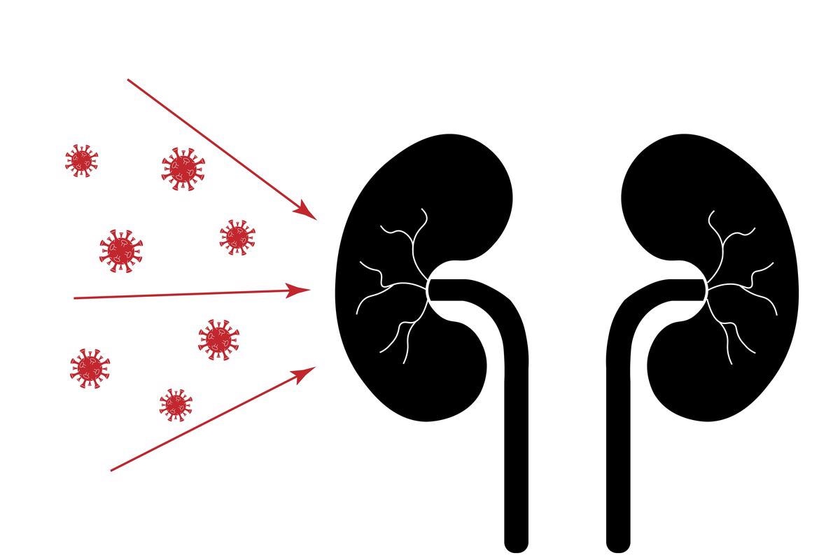
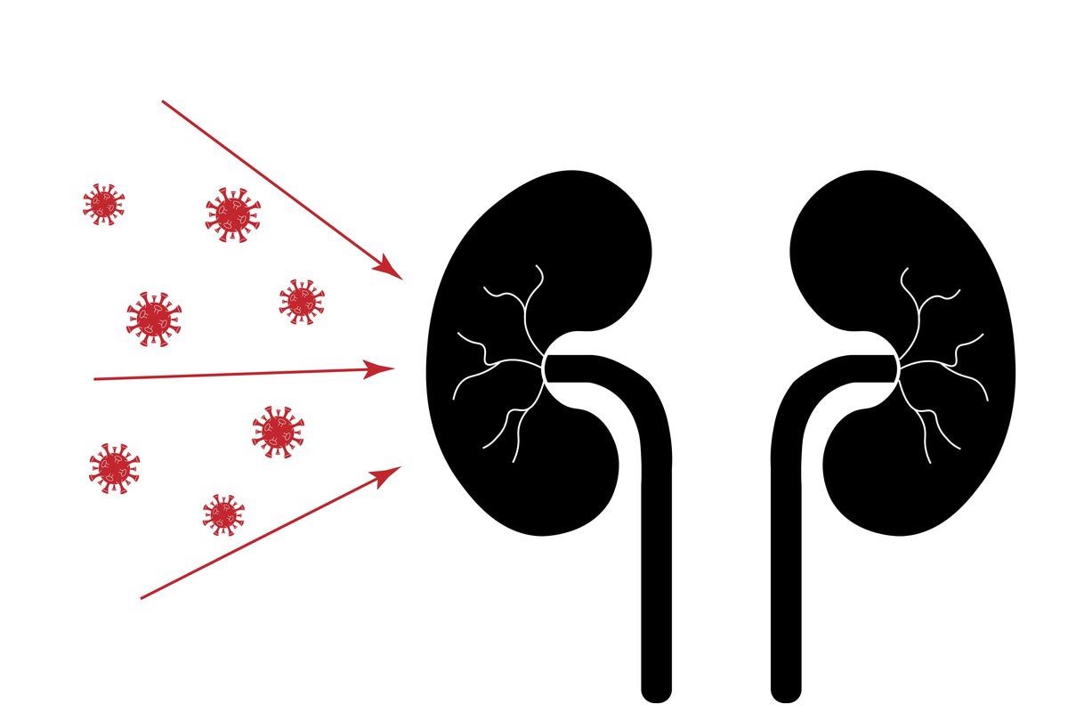
Coronavirus disease 2019 (COVID-19) is caused by the severe acute respiratory syndrome coronavirus 2 (SARS-CoV-2), which is the causative virus of the current global pandemic. Research suggests that SARS-CoV-2 infection affects organs other than the lungs despite its initial manifestation as pulmonary disease. The clinical picture of COVID-19 has been improved thanks to autopsy research, case reports, and retrospective clinical studies. SARS-CoV-2 has been linked to various diseases, including neurological symptoms, increased risk of acute-on-chronic liver failure, altered hemostasis leading to venous thromboembolism, and acute kidney injury (AKI).
 Study: SARS-CoV-2 infects the human kidney and drives fibrosis in kidney organoids. Image Credit: Eugene B-sov/Shutterstock
Several recent reports show that COVID-19 patients have an infection of various renal cell types, including proximal tubule (PT) epithelial cells. However, it is currently unknown whether or not this infection has pathophysiologic implications for the kidney. SARS-CoV-2 directly infects and destroys kidney cells, according to new findings, albeit the molecular alterations associated with this infection are unknown.
AKI in COVID-19 individuals could be a severe complication of acute respiratory distress syndrome (ARDS) requiring critical care, a result of direct infection of kidney cells, or a combination of the two. SARS-CoV-2 infects podocytes and tubular epithelium in human kidneys, causing kidney damage and fibrosis, which could explain the development of chronic kidney disease (CKD) following COVID-19.
Researchers infected human-induced pluripotent-stem-cell (iPSC)-derived kidney organoids with SARS-CoV-2 to study the virus's direct effect on the kidney, taking advantage of the model system's ability to rule out systemic effects of the disease or renal effects of intensive care medical treatment. SARS-CoV-2 infection induces cellular damage and activation of numerous profibrotic signaling pathways, according to the findings, which are mediated by cellular crosstalk between infected epithelium and podocytes and interstitial fibroblasts. The researchers reported their study in the journal Cell Stem Cell.
The study
The authors obtained kidney tissue from 62 COVID-19 patients to investigate the effects of SARS-CoV-2 on the kidney. The SARS-CoV-2 nucleocapsid protein was found in the cytoplasm of proximal tubular (PT) epithelium expressing angiotensin-converting enzyme 2 (ACE2), consistent with prior studies.
For the nucleocapsid protein staining, SARS-CoV-2 infected lung tissue was used as a positive control, and non-COVID-19 autopsy and nephrectomy tissues were used as negative controls. Kidney injury molecule 1 (KIM1) expression in the biopsy specimen's lotus tetragonolobus lectin (LTL) positive tubules revealed PT injury. KIM1 expression was also found in some PTs of COVID-19 patient autopsy renal tissue.
The kidney's response to injury, regardless of the type, eventually leads to fibrosis, which is the characteristic of CKD. The scientists found that COVID-19 patients had more interstitial fibrosis in their kidneys than a control group matched for age, sex, and comorbidities. COVID-19 patients' kidneys had considerably higher quantities of extracellular matrix (ECM) than the control cohort, according to Masson's Trichrome staining and quantitative collagen expression in kidney tissue of all patients.
The authors used single-nucleus RNA sequencing (snRNA-seq) of autopsy kidney tissue recovered from a COVID-19 patient in the cohort to validate their hypothesis that SARS-CoV-2 may directly enter and infect kidney cells. From a recently publicly available kidney snRNA-seq dataset, snRNA-seq showed 13 different cell clusters, which were annotated by label transfer. The authors used a targeted sequencing strategy combined with a typical 10X Genomics workflow to track down SARS-CoV-2 in the dataset.
The authors computationally linked the SARS-CoV-2 readings back to individual cells after sequencing, allowing them to track viral RNA expression in nearly all cell clusters. Due to the sparsity of the 3′ sequencing, several SARS-CoV-2 entry factors were not found in the dataset. However, the researchers discovered overexpression of the SARS-CoV-2 infection-related genes PLCG2 and AFDN. Increased collagen, ECM glycoprotein, and proteoglycan expression were detected in COVID-19 kidney tissue fibroblasts when extracellular matrix (ECM) remodeling was studied using gene set enrichment analysis (GSEA).
The authors used human iPSC-derived kidney organoids to replicate SARS-CoV-2 infection in vitro to explore the virus's direct impact on kidney cells independent of systemic effects. An altered version of the widely used Little technique was utilized to create kidney organoids. Human-induced pluripotent-stem-cell (iPSCs) briefly differentiated into ureteric epithelium or metanephric mesenchyme lineages by exposing them to the WNT agonist CHIR 99021 for three days or five days. To activate morphogenic signals necessary for segmented patterning, cells were combined in a 1:2 ratio on day 7 to induce morphogenic cues.
Before SARS-CoV-2 infection, organoids were grown in a 3D air-liquid interface transwell system until day 7+18. Immunofluorescence staining revealed the presence of nephron-like structures in the kidney organoids; as evidenced by nephrin (podocytes), Lotus tetragonolobus lectin (LTL, PTs) co-stained with ACE2, which is expressed at the apical side of the proximal tubular cells, similar to human kidneys, E-cadherin, and E-cadherin plus GATA3 On day 7+18, organoids were injected with SARS-CoV-2 viral particles. The presence of viral RNA in organoids verified the infection. Using in situ hybridization, the authors validated SARS-CoV-2 mRNA expression in podocytes and proximal tubular epithelium.
Implications
Taken together, these findings reveal that SARS-CoV-2 infection of kidney cells resulted in a molecular switch to profibrotic signaling and tissue damage. The data supports a concept in which SARS-CoV-2 kidney injury is partially independent of systemic consequences of the disease or intensive care treatment and instead represents a direct effect of the virus because organoids lack immune cells and perfusion. As a result, a viral infection could cause acute damage and fibrotic remodeling, leading to renal dysfunction and CKD.
-
Jansen, J. et al. (2021) "SARS-CoV-2 infects the human kidney and drives fibrosis in kidney organoids", Cell Stem Cell. doi: 10.1016/j.stem.2021.12.010. https://www.sciencedirect.com/science/article/pii/S1934590921005208?via%3Dihub
Posted in: Medical Science News | Medical Research News | Disease/Infection News
Tags: ACE2, Acute Kidney Injury, Acute Respiratory Distress Syndrome, Angiotensin, Angiotensin-Converting Enzyme 2, Biopsy, Cadherin, Cell, Chronic, Chronic Kidney Disease, Collagen, Coronavirus, Coronavirus Disease COVID-19, covid-19, Critical Care, Cytoplasm, Enzyme, Fibrosis, Gene, Genes, Genomics, Glycoprotein, Hemostasis, Hybridization, in vitro, Intensive Care, Kidney, Kidney Disease, Liver, Lungs, Mesenchyme, Molecule, Nephrectomy, Organoids, Pandemic, Protein, Proteoglycan, Research, Respiratory, RNA, RNA Sequencing, SARS, SARS-CoV-2, Severe Acute Respiratory, Severe Acute Respiratory Syndrome, Syndrome, Thromboembolism, Venous Thromboembolism, Virus
.jpg)
Written by
Colin Lightfoot
Colin graduated from the University of Chester with a B.Sc. in Biomedical Science in 2020. Since completing his undergraduate degree, he worked for NHS England as an Associate Practitioner, responsible for testing inpatients for COVID-19 on admission.
Source: Read Full Article
