Brain activity during psychological stress may predict chest pain in people with heart disease
Stress-induced activity in the inferior frontal lobe of the brain may have a direct correlation with chest pain among people with coronary artery disease, according to new research released today in Circulation: Cardiovascular Imaging.
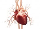
Angina pectoris, or stable angina, is chest pain or discomfort due to inadequate blood flow to the heart. Angina occurs due to coronary artery disease, and, while recent research has indicated psychological factors including mental stress can lead to angina, little is known about the brain mechanisms involved. This study was designed to measure how activity in the inferior frontal lobe of the brain—the area of the brain associated with emotional regulation and stress—affects the severity of self-reported angina.
“Our study sought to understand the degree to which health care providers should incorporate stress and other psychological factors when evaluating and treating angina,” said lead investigator Amit J. Shah, M.D., M.S.C.R., assistant professor of epidemiology at Emory University’s Rollins School of Public Health in Atlanta. “Although brain imaging during a mental stress challenge is not a test that can be ordered in clinical settings, the study shows an important proof-of-concept that shows the brain’s reactivity to stress is an important consideration when considering angina treatment.”
A total of 148 people with coronary artery disease participated in the study from 2011 to 2014. The study participants were, on average, 62 years old, and consisted of 69% men and 31% women. The group underwent testing of mentally stressful events in a clinical setting and had brain imaging and cardiac imaging conducted in conjunction with the tests.
Participants were assessed with three tests that were performed over a two-week period: a mental stress test with brain imaging; a mental stress test with heart imaging; and an exercise or chemical stress test with heart imaging. During these tests, the researchers monitored participants for chest pain. Additional questionnaires for chest pain and cardiovascular events were assessed after two years.
Investigators examined factors related to the severity of the participants’ angina and observed that brain activity in the inferior frontal lobe showed the strongest relationship with self-reported angina at baseline and also at a two-year follow-up appointment. The results indicated that:
- participants who reported having monthly, weekly or daily angina symptoms had higher inferior frontal lobe activity in response to mental stress at both baseline;
- those who reported angina during mental stress testing with cardiac imaging also had higher inferior frontal lobe activation compared to individuals who did not have active chest pain during mental stress testing; and
- there was a significant association between inferior frontal lobe activation during stress and the degree of change in angina frequency at the two-year follow-up, suggesting that brain-related changes might predict worsened future angina.
“We were surprised by the strength of the relationship between the level of activity in this brain region and the frequency of chest pain reported, as well as the lack of a relationship to factors that are normally considered important when treating angina, such as heart imaging,” said Shah. “The top three factors that explained angina frequency were all stress related, including brain activation, depressive symptoms and PTSD symptoms. This is surprising because when we manage angina in clinical settings, we normally do not consider stress as an underlying factor, and rather focus on blood flow in the heart.”
Source: Read Full Article