Antibody Purification Techniques
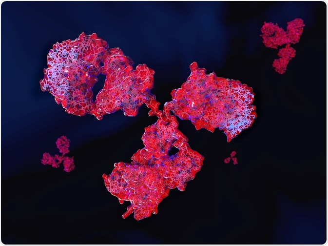
Many diagnostic technologies rely on the detection of antigens by antibodies. An example of this is the molecular biology technique, ELISA.
 Image Credit: Juan Gaertner / Shutterstock
The antibodies used in diagnostic techniques are often generated in vivo, when an animal is injected with a specific antigen from another species. The animal's immune system responds by producing antibodies, which can be harvested by taking a serum sample.
This “antiserum” isolated from the animal is then subjected to a purification step to remove contaminants, or to obtain just one type of antibody (monoclonal) from a mixture of antibodies (polyclonal).
Physicochemical fractionation
Physicochemical fractionation describes methods that separate antibodies by size, charge, or chemical properties. The different classes of immunoglobulin (Ig), such as IgM and IgG, often have similar amino acid compositions, solubility, and structure. Physicochemical fractionation exploits this, by removing any molecule that does not carry the same properties.
Size exclusion chromatography
In this method, the molecules are sorted based on their size and molecular weight. The chromatography column consists of dextran, agarose, or polyacrylamide beads. Based on the size of these beads, different size of macromolecules can be isolated.
The low molecular weight components can be removed by dialysis, desalting, and diafiltration. Then, size exclusion resins that consist of high molecular cut-off can further separate immunoglobulins that are less than 140 kDa.
Ammonium sulphate precipitation
This method is used to isolate antibodies from serum, cell culture supernatant, or ascites fluid. The increasing concentration of ammonium sulphate makes proteins and other macromolecules increasingly insoluble, leading to their precipitation.
Different proteins, including antibodies, precipitate at specific concentrations of ammonium sulphate. For example, antibodies precipitate at 40-50% ammonium sulphate. The purity of antibodies precipitated using this method depends on the temperature, pH, the rate at which salt is added, and time.
Ion exchange chromatography
Ion exchange chromatography is used to isolate or separate compounds based on their charge. The column consists of positively or negatively charged resins which then bind to negatively or positively charged proteins.
The buffer system can be modified so that the antibody of interest is bound and released in a highly specific way. Alternatively, the system can be designed to bind everything other than the target antibody. This method is cost effective to purify antibodies.
Immobilized metal chelate chromatography
In this method, chelate immobilized divalent metal ions are used to bind proteins that contain three or more histidine residues.
Mammalian IgGs consist of histidine clusters, which can therefore bind to immobilized nickel on a chromatography column. Thus, this method can be used to purify and isolate IgGs from a mixture.
Purifying antibodies based on their classification
Antibodies have evolutionarily conserved structures. Several methods have been developed which isolate antibodies based on the similarities of each class.
Protein A, G, and L
Protein A, G, and L are bacterial proteins and they have specific domains which bind with specific immunoglobulins. Each of the proteins have specific binding properties; protein A and G bind to the Fc region, while protein L bind to the light chain of the Fab region.
Protein A does not bind to IgD and only weakly binds to IgA and IgM. Protein G only binds to IgG, while protein L binds to all antibody classes. Thus, these bacterial proteins can be used to isolate a specific class of immunoglobulin. For example, in a chromatography column with protein A, serum can be passes through the column and the protein A is able to bind to and separate IgG from the serum.
Purification of IgM
Proteins G and A do not bind strongly with IgM as there is steric hindrance of the binding regions on IgM. Thus, to purification of IgM involves several methods, including ammonium sulfate precipitation, ion exchange chromatography, gel filtration, and zone electrophoresis.
Purification of IgA
This method was uncovered when a D-galactose lectin called jacalin was extracted from jackfruit seeds. This compound was found to contain four identical domains, and it binds to IgA. Jacalin can be immobilized on agarose gels and subsequently can be used to purify and separate IgA from other immunoglobulins.
Purification of IgY
IgY is a unique immunoglobulin made by chickens, and it is present in high quantities in egg yolk. Proteins A, G, and L cannot be used to purify IgY as these proteins do not bind to IgY. Thus, ammonium sulfate precipitation method is used to purify IgY.
Antigen-specific affinity purification of antibodies
As antibodies bind to a specific antigen, this interaction can also be used to purify antibodies. Affinity purification involves immobilizing an antigen which is specific for an antibody, such that only antibodies which bind to the specific antigen are isolated. This method is called ligand immobilization method for affinity purification.
Sources
- Antibody Purification Methods by Thermo Fisher.
- Antibody Purification Methods by Sino Biological.
- An Education in Antibody Purification Methods.
Further Reading
- All Antibody Content
- Antibody – What is an Antibody?
- Antibody Forms
- Antibodies in Medicine
- Antibody Structure
Last Updated: Oct 22, 2018

Written by
Dr. Surat P
Dr. Surat graduated with a Ph.D. in Cell Biology and Mechanobiology from the Tata Institute of Fundamental Research (Mumbai, India) in 2016. Prior to her Ph.D., Surat studied for a Bachelor of Science (B.Sc.) degree in Zoology, during which she was the recipient of anIndian Academy of SciencesSummer Fellowship to study the proteins involved in AIDs. She produces feature articles on a wide range of topics, such as medical ethics, data manipulation, pseudoscience and superstition, education, and human evolution. She is passionate about science communication and writes articles covering all areas of the life sciences.
Source: Read Full Article


